|
ATCC
glycerol 8 hl 60 Glycerol 8 Hl 60, supplied by ATCC, used in various techniques. Bioz Stars score: 99/100, based on 1 PubMed citations. ZERO BIAS - scores, article reviews, protocol conditions and more https://www.bioz.com/result/glycerol 8 hl 60/product/ATCC Average 99 stars, based on 1 article reviews
glycerol 8 hl 60 - by Bioz Stars,
2026-03
99/100 stars
|
Buy from Supplier |
|
Thermo Fisher
rpmi 1640 Rpmi 1640, supplied by Thermo Fisher, used in various techniques. Bioz Stars score: 99/100, based on 1 PubMed citations. ZERO BIAS - scores, article reviews, protocol conditions and more https://www.bioz.com/result/rpmi 1640/product/Thermo Fisher Average 99 stars, based on 1 article reviews
rpmi 1640 - by Bioz Stars,
2026-03
99/100 stars
|
Buy from Supplier |
|
DSMZ
primary aml cells hl 60 Primary Aml Cells Hl 60, supplied by DSMZ, used in various techniques. Bioz Stars score: 96/100, based on 1 PubMed citations. ZERO BIAS - scores, article reviews, protocol conditions and more https://www.bioz.com/result/primary aml cells hl 60/product/DSMZ Average 96 stars, based on 1 article reviews
primary aml cells hl 60 - by Bioz Stars,
2026-03
96/100 stars
|
Buy from Supplier |
|
National Centre for Cell Science
human epidermoid carcinoma (a431) cell line  Human Epidermoid Carcinoma (A431) Cell Line, supplied by National Centre for Cell Science, used in various techniques. Bioz Stars score: 90/100, based on 1 PubMed citations. ZERO BIAS - scores, article reviews, protocol conditions and more https://www.bioz.com/result/human epidermoid carcinoma (a431) cell line/product/National Centre for Cell Science Average 90 stars, based on 1 article reviews
human epidermoid carcinoma (a431) cell line - by Bioz Stars,
2026-03
90/100 stars
|
Buy from Supplier |
|
ATCC
2018 leukemia ccrf cem 109 53 2018 Leukemia Ccrf Cem 109 53, supplied by ATCC, used in various techniques. Bioz Stars score: 97/100, based on 1 PubMed citations. ZERO BIAS - scores, article reviews, protocol conditions and more https://www.bioz.com/result/2018 leukemia ccrf cem 109 53/product/ATCC Average 97 stars, based on 1 article reviews
2018 leukemia ccrf cem 109 53 - by Bioz Stars,
2026-03
97/100 stars
|
Buy from Supplier |
|
ATCC
u 87mg atcc hl 60  U 87mg Atcc Hl 60, supplied by ATCC, used in various techniques. Bioz Stars score: 99/100, based on 1 PubMed citations. ZERO BIAS - scores, article reviews, protocol conditions and more https://www.bioz.com/result/u 87mg atcc hl 60/product/ATCC Average 99 stars, based on 1 article reviews
u 87mg atcc hl 60 - by Bioz Stars,
2026-03
99/100 stars
|
Buy from Supplier |
|
ATCC
hl60 mx2 cells Hl60 Mx2 Cells, supplied by ATCC, used in various techniques. Bioz Stars score: 93/100, based on 1 PubMed citations. ZERO BIAS - scores, article reviews, protocol conditions and more https://www.bioz.com/result/hl60 mx2 cells/product/ATCC Average 93 stars, based on 1 article reviews
hl60 mx2 cells - by Bioz Stars,
2026-03
93/100 stars
|
Buy from Supplier |
|
Proteintech
pad4 primary antibodies  Pad4 Primary Antibodies, supplied by Proteintech, used in various techniques. Bioz Stars score: 95/100, based on 1 PubMed citations. ZERO BIAS - scores, article reviews, protocol conditions and more https://www.bioz.com/result/pad4 primary antibodies/product/Proteintech Average 95 stars, based on 1 article reviews
pad4 primary antibodies - by Bioz Stars,
2026-03
95/100 stars
|
Buy from Supplier |
|
ATCC
u937  U937, supplied by ATCC, used in various techniques. Bioz Stars score: 99/100, based on 1 PubMed citations. ZERO BIAS - scores, article reviews, protocol conditions and more https://www.bioz.com/result/u937/product/ATCC Average 99 stars, based on 1 article reviews
u937 - by Bioz Stars,
2026-03
99/100 stars
|
Buy from Supplier |
|
Millipore
human promyelocytic leukemia hl60 cells  Human Promyelocytic Leukemia Hl60 Cells, supplied by Millipore, used in various techniques. Bioz Stars score: 90/100, based on 1 PubMed citations. ZERO BIAS - scores, article reviews, protocol conditions and more https://www.bioz.com/result/human promyelocytic leukemia hl60 cells/product/Millipore Average 90 stars, based on 1 article reviews
human promyelocytic leukemia hl60 cells - by Bioz Stars,
2026-03
90/100 stars
|
Buy from Supplier |
|
ATCC
tm hl 60 Tm Hl 60, supplied by ATCC, used in various techniques. Bioz Stars score: 94/100, based on 1 PubMed citations. ZERO BIAS - scores, article reviews, protocol conditions and more https://www.bioz.com/result/tm hl 60/product/ATCC Average 94 stars, based on 1 article reviews
tm hl 60 - by Bioz Stars,
2026-03
94/100 stars
|
Buy from Supplier |
|
Inserm Transfert
fpr1-hl60 cells Fpr1 Hl60 Cells, supplied by Inserm Transfert, used in various techniques. Bioz Stars score: 90/100, based on 1 PubMed citations. ZERO BIAS - scores, article reviews, protocol conditions and more https://www.bioz.com/result/fpr1-hl60 cells/product/Inserm Transfert Average 90 stars, based on 1 article reviews
fpr1-hl60 cells - by Bioz Stars,
2026-03
90/100 stars
|
Buy from Supplier |
Image Search Results
Journal: ACS Omega
Article Title: Cytotoxic and Apoptotic Effects of Chemogenic and Biogenic Nano-sulfur on Human Carcinoma Cells: A Comparative Study
doi: 10.1021/acsomega.1c04047
Figure Lengend Snippet: Morphological changes of A549, A431, HL60, and IMR90 cell-lines un-treated (a, e, i, m) and, treated with IC 50 concentration of the standard drug (b, f, j, n), SNP-B (c, g, k, o), and SNP-C (d, h, l, p) after 24 h treatment.
Article Snippet: Human epidermoid carcinoma (A431),
Techniques: Concentration Assay

Journal: ACS Omega
Article Title: Cytotoxic and Apoptotic Effects of Chemogenic and Biogenic Nano-sulfur on Human Carcinoma Cells: A Comparative Study
doi: 10.1021/acsomega.1c04047
Figure Lengend Snippet: Apoptosis of A549, A431, HL60, and IMR90 cell-lines un-treated (a, e, i, m) and, treated with IC 50 concentration of the standard drug (b, f, j, n), SNP-B (c, g, k, o), and SNP-C (d, h, l, p).
Article Snippet: Human epidermoid carcinoma (A431),
Techniques: Concentration Assay

Journal: ACS Omega
Article Title: Cytotoxic and Apoptotic Effects of Chemogenic and Biogenic Nano-sulfur on Human Carcinoma Cells: A Comparative Study
doi: 10.1021/acsomega.1c04047
Figure Lengend Snippet: Cell cycle analysis of A549, A431, HL60, and IMR90 cell lines untreated (a, e, i, m) and, treated with IC 50 concentration of the standard drug (b, f, j, n), SNP-B (c, g, k, o), and SNP-C (d, h, l, p).
Article Snippet: Human epidermoid carcinoma (A431),
Techniques: Cell Cycle Assay, Concentration Assay

Journal: ACS Omega
Article Title: Cytotoxic and Apoptotic Effects of Chemogenic and Biogenic Nano-sulfur on Human Carcinoma Cells: A Comparative Study
doi: 10.1021/acsomega.1c04047
Figure Lengend Snippet: Caspase-3 expression of A549, A431, HL60, and IMR90 cell-lines un-treated (a, e, i, m), treated with IC 50 concentration of the standard drug (b, f, j, n), SNP-B (c, g, k, o), and SNP-C (d, h, l, p).
Article Snippet: Human epidermoid carcinoma (A431),
Techniques: Expressing, Concentration Assay
 a " width="100%" height="100%">
Journal: ACS Omega
Article Title: Cytotoxic and Apoptotic Effects of Chemogenic and Biogenic Nano-sulfur on Human Carcinoma Cells: A Comparative Study
doi: 10.1021/acsomega.1c04047
Figure Lengend Snippet: Percentage Cell Viability of A549, A431, HL60 and, IMR90 Cell-Lines Treated with Various Concentrations of SNP-B and SNP-C
Article Snippet: Human epidermoid carcinoma (A431),
Techniques: Control
 a " width="100%" height="100%">
Journal: ACS Omega
Article Title: Cytotoxic and Apoptotic Effects of Chemogenic and Biogenic Nano-sulfur on Human Carcinoma Cells: A Comparative Study
doi: 10.1021/acsomega.1c04047
Figure Lengend Snippet: Apoptotic Study (i.e., Annexin V/PI Expression Study) of SNP-B and SNP-C against A549, A431, HL60, and IMR90 Cell-Lines
Article Snippet: Human epidermoid carcinoma (A431),
Techniques: Expressing
 a " width="100%" height="100%">
Journal: ACS Omega
Article Title: Cytotoxic and Apoptotic Effects of Chemogenic and Biogenic Nano-sulfur on Human Carcinoma Cells: A Comparative Study
doi: 10.1021/acsomega.1c04047
Figure Lengend Snippet: Cell Cycle Analysis of SNP-B and SNP-C against A549, A431 HL60, and IMR90 Cell Lines
Article Snippet: Human epidermoid carcinoma (A431),
Techniques: Cell Cycle Assay
 a " width="100%" height="100%">
Journal: ACS Omega
Article Title: Cytotoxic and Apoptotic Effects of Chemogenic and Biogenic Nano-sulfur on Human Carcinoma Cells: A Comparative Study
doi: 10.1021/acsomega.1c04047
Figure Lengend Snippet: Caspase-3 Expression Study of SNP-B and SNP-C against A549, A431, HL60, and IMR90 Cell Lines
Article Snippet: Human epidermoid carcinoma (A431),
Techniques: Expressing, Fluorescence
Journal: Biomedicines
Article Title: The Thioredoxin System of Mammalian Cells and Its Modulators
doi: 10.3390/biomedicines10071757
Figure Lengend Snippet: Cytotoxicity of TrxR inhibitors.
Article Snippet: 3. Organoselenium compounds , 3.1. Organo- selenium compounds 3.2. Phenylarsenic oxide derivatives (dithiarsanes) , 1,2-(5,5′-Dimethoxy(bis-1,2-benzisoselenazol-3(2 H )-one))ethane 2-(4-Aminophenyl)-1,3,2-dithiarsenane (PAO−PDT) , , 1.64 0.6 ,
Techniques: Analogues
Journal: bioRxiv
Article Title: Citrullination of TDP-43 is a key post-translation modification associated with structural and functional changes and progressive pathology in TDP-43 mouse models and human proteinopathies
doi: 10.1101/2025.02.28.639952
Figure Lengend Snippet: a, The enzymatic reaction mediated by PADs converts arginine to citrulline. b, c, Western blot images of PAD2 and PAD4-mediated citrullination of H3 (b) and TDP-43 (c). In (c) arrows indicate the observed shift in TDP-43 molecular weight. d, Coomassie staining of PAD2 (bands 1, 4), PAD4 (bands 2, 6), unmodified TDP-43 (band 3), PAD2-mediated citR TDP-43 (band 5), and PAD4-mediated citR TDP-43 (band 7). e, Bar diagrams showing the MW shift (kDa) of TDP-43 protein following citrullination. f, Schematic representation of arginine epitopes positioned in the human TDP-43 protein sequence. Position of the 11 citrullinated arginine epitopes are indicated in red. g, Six out of eleven arginine epitopes susceptible to citrullination laid within common RX(X)R (red box) or RXG/RGGG (green box) motifs in TDP-43 sequence. h, MS/MS spectrum showing b- and y-ion coverage of modified citR83 peptide and extracted ion chromatograms (XICs) showed abundance and retention time of unmodified R83 vs. citR83 with PAD2 and PAD4 treatment (intact peptide monoisotopic m/z (+2) 719.8457, (+3) 480.2329). i, Retention time peaks for TDP-43 peptides surrounding the unmodified or modified R83 epitope and base peak m/z undergoing methionine (Met85) oxidation, citrullination or both showed reliable time separation.
Article Snippet: PAD2 and
Techniques: Western Blot, Molecular Weight, Staining, Sequencing, Tandem Mass Spectroscopy, Modification

Journal: bioRxiv
Article Title: Citrullination of TDP-43 is a key post-translation modification associated with structural and functional changes and progressive pathology in TDP-43 mouse models and human proteinopathies
doi: 10.1101/2025.02.28.639952
Figure Lengend Snippet: Spectra showing b- and y-ion coverage of citrullinated peptides, as well as XICs retention times for unmodified and citrullinated peptides treated with PAD2 and PAD4, corresponding to a - c, citR165 (intact peptide monoisotopic m/z (+2) 665.8214), d - f, citR191(intact peptide monoisotopic m/z (+2) 672.8469), g - i, citR268/272 (intact peptide monoisotopic m/z (+2) 728.3560), j - l, citR293 (intact peptide monoisotopic m/z (+2) 638.29).
Article Snippet: PAD2 and
Techniques:

Journal: bioRxiv
Article Title: Citrullination of TDP-43 is a key post-translation modification associated with structural and functional changes and progressive pathology in TDP-43 mouse models and human proteinopathies
doi: 10.1101/2025.02.28.639952
Figure Lengend Snippet: a, Schematic representation of seven polyclonal antibodies raised against the citrullinated arginine epitopes. Epitope positions within each TDP-43 functional domain is indicated. b, Antibody specificity was determined by western blot via PAD4-mediated citrullinated TDP-43 protein probed with citR83 (NLS), citR165 and citR191 (RRM1, RRM2), citR268/272, citR275 (CTD) antibodies. c, Immunohistochemical images of TAR4/4 and non-Tg littermates cortical tissue labeled against citR TDP-43 antibody panel. Increased neuronal expression of citR83, citR165, citR191, citR268/272 and citR275 in TAR4/4 tissue compared to Non-Tg tissue. Arrows indicate the citR TDP-43 morphologies observed with the citR268/272 and citR275 antibodies raised against TDP-43 C-terminal domain (arrows). Insets represent magnified neuronal citR TDP-43 morphological properties recognized from each antibody. d, Images of cortical tissue incubated with secondary antibody only - “no-primary” control. n = 3, scale bar is 50µm and 200 µm.
Article Snippet: PAD2 and
Techniques: Functional Assay, Western Blot, Immunohistochemical staining, Labeling, Expressing, Incubation, Control
Journal: bioRxiv
Article Title: Citrullination of TDP-43 is a key post-translation modification associated with structural and functional changes and progressive pathology in TDP-43 mouse models and human proteinopathies
doi: 10.1101/2025.02.28.639952
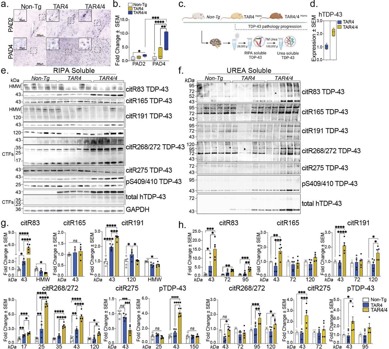
Figure Lengend Snippet: a, Immunohistochemical images of Non-Tg, TAR4 and TAR4/4 cortex labeled with PAD2 and PAD4 antibodies. b, Fold change of PAD2 and PAD4 (% area mean of the values ± SEM) normalized to the Non-Tg control. One-way or Two-way ANOVA, followed by Tukey’s post hoc multiple comparisons tests, n = 5, *p < 0.05, **p < 0.01, ***p < 0.001, ****p < 0.0001. c, Schematic overview of the TAR TDP-43 mouse model and the fractionation of cortical tissue into RIPA-soluble and (7 M) Urea-soluble fractions. d, The expression levels of the human TDP-43 in the TAR model. e, Cortical RIPA soluble and f, Urea soluble fraction analyzed by Western blotting and probed for citR TDP-43 antibody panel (citR83, 165, 191, 268/272, 275), pTDP-43 409/410 and total human TDP-43 protein. g, Quantification of citR TDP-43 43 kDa protein levels, proteolytic fragments (17, 25 & 35 kDa) and intermediate high molecular species (72, 98 & 120k Da) normalized to GAPDH in RIPA and h, Urea fraction normalized to the total protein loaded signal. Data represent the mean of the values ± SEM; One-way ANOVA, followed by Tukey’s or Šidák post hoc multiple comparisons tests, n = 5, *p < 0.05, **p < 0.01, ***p < 0.001, ****p < 0.0001.
Article Snippet: PAD2 and
Techniques: Immunohistochemical staining, Labeling, Control, Fractionation, Expressing, Western Blot
Journal: Immunology
Article Title: Functional exhaustion of CD4 + T cells induced by co‐stimulatory signals from myeloid leukaemia cells
doi: 10.1111/imm.12665
Figure Lengend Snippet: Up‐regulation of surface molecules related to activation and/or exhaustion on CD4 + T cells co‐cultured with different myeloid leukaemia cell lines for 96 hr under continuous stimuli conditions
Article Snippet: Human myeloid leukaemia cell lines, KG‐1, Kasumi‐1, HL‐60,
Techniques: Activation Assay, Expressing
Journal: The Journal of Biological Chemistry
Article Title: Mitigation of NADPH Oxidase 2 Activity as a Strategy to Inhibit Peroxynitrite Formation
doi: 10.1074/jbc.M115.702787
Figure Lengend Snippet: Step-by-step protocol applied to screen the library of bioactive compounds at The Broad Institute
Article Snippet:
Techniques: Cell Culture, Concentration Assay, Inhibition, Fluorescence

Journal: The Journal of Biological Chemistry
Article Title: Mitigation of NADPH Oxidase 2 Activity as a Strategy to Inhibit Peroxynitrite Formation
doi: 10.1074/jbc.M115.702787
Figure Lengend Snippet: Positive hits obtained during screening of the library of bioactive compounds using three probes as follows: hydropropidine (50 μ m ) in the presence of DNA (0.1 mg/ml) as a probe for Ȱ, and coumarin boronic acid (100 μ m ) or Amplex Red (50 μ m ) in the presence of HRP (0.1 units/ml) as probes for H 2 O 2 Differentiated HL60 cells were stimulated with PMA (1 μ m ) to induce Nox2 activity in HBSS supplemented with HEPES buffer (25 m m ) and dtpa (0.1 m m ).
Article Snippet:
Techniques: Activity Assay

Journal: The Journal of Biological Chemistry
Article Title: Mitigation of NADPH Oxidase 2 Activity as a Strategy to Inhibit Peroxynitrite Formation
doi: 10.1074/jbc.M115.702787
Figure Lengend Snippet: IC 50 values for selected positive hits, determined by monitoring the effect of the compounds on the rate of oxidation of coumarin boronic acid (100 μ m ) as a probe for H 2 O 2 Differentiated HL60 cells were stimulated with PMA (1 μ m ) to induce Nox2 activity in HBSS supplemented with HEPES buffer (25 m m ) and dtpa (0.1 m m ) and the extent of probe oxidation was monitored in 384-well plates using fluorescence plate reader.
Article Snippet:
Techniques: Activity Assay, Fluorescence